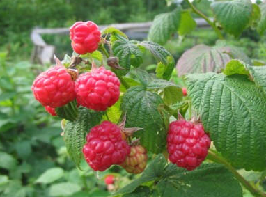
Pesticide Detection Methods Development
Experiment 3 (Finished on 3/2/14)
Purpose:
Estimate the Molar Absorptivity Coefficient (e) of the measurable component of the pesticide detection (cholinesterase inhibition) assay used for testing organic and conventional raspberries.
Procedure:
Generate a serial dilution of the control solution using the following schedule (fig.1) and measure the absorbance of each dilution at each step of the dilution:
fig.1 serial dilution schedule
Results:
Observed that the there is a small amount of measurable absorbance in the substrate (Solution 2) at high concentrations. There there is no measurable absorbance when the substrate is mixed with Cholinesterase. Once the mixture of substrate and enzyme is buffered with Solution 3 (sodium acetate) and the pH is stabilized, there is a measurable absorbance for mixtures that are at least 25% stock solution (Solution 1 + 2 + 3 + 5min incubation). Figure 2 show that there we can extract a Molar Absorptivity Coefficient from the region between 25% to 100% stock solution.
fig 2. absorbance for various dilutions of the assay at various stages of preparation.
Figure 3 shows how the Molar Absorptivity Coefficient is calculated for stock dilutions between 25% to 100% (the dilutions were converted to Cholinesterase concentration (M)). Based on the following formula:
A = ecl
Where A = Absorbance @ 415nm, e = Molar Absorptivity Coefficient (M-1 cm-1), c = concentration (M), l = pathlength (1 cm for cuvettes used).
Molar Absorptivity Coefficient (e) = A/cl = 2865/M*cm
fig 3. absorbance for 25% to 100% stock dilutions, converted to M for Molar Absorptivity Coefficient calculations.
Conclusion:
The assay appears to be very sensitive to pH, which is expected for any type of biochemical system. As we were not able to measure the absorbance of substrate or substrate + enzyme, its clear that enzyme will not start to convert substrate until the system has a buffer in place to stabilize the pH (which needs to be measured). For the above calculations, we are assuming a 1-to-1 conversion between substrate & enzyme. But, this may not be the case and should be investigated further.
///////////////////////////////////////////////////////////////////////////////////////////////////////////////////////////////////////////////
Experiment 2 (Finished on 2/27/14)
Purpose:
Compare a methanol extraction to the results from using the extraction solution provided by the kit manufacturer. Use Raspberries from the same batch and run with a control.
Procedure:
Samples: Driscoll's Organic Raspberries (purchased at Costco in Mountain View, CA). I used the website mydriscolls.com and got specific information about the farm, and its located in Oxnard, CA. Driscolls now has a link on this portal to something called HarvestMark. I followed it and entered the barcode information and got the following information:
Bar Code: 5997 4716 5149 DS08
I don't know what Ranch ID and UPC mean, but I will look into it and see if I can find out more information.
Methods used are the same as those outline in my original post with regards to sample prep: http://publiclab.org/notes/silverhammer/02-06-2014/detecting-pesticides-in-organic-and-conventional-raspberries-using-open-source-instrumentation
To better control the experiment, I will use 6 raspberries from the same container, and then each berry will be cut in half and then one half will be used for the methanol extraction, while the other half will be used with the kit extraction solvent (dichloromethane).
The evaporation of methanol will take longer at the water temperatures used for the kit extraction solvent, so the water temperature will brought to ~77C to ensure that the methanol evaporates.
Samples will be monitored at 1min intervals on the PublicLab spectrometer as well as the IO Rodeo Colorimeter with a 415nm LED. PublicLab spectrometer setup will be 9" pathlength, 40W fluorescent backlight.
Results
The extract solutions look very different from each other after the extraction. The kit extract solution has a slight pink color to it, but otherwise is fairly clear. The methanol extract takes on a very dark red color.
The extraction process using methanol proved to be more challenging than expected. The boiling point for methanol is ~77C. After 30 minutes of evaporation at ~77C, there was still 0.5ml of dark red solution at the bottom of the test tube. I was not able to completely evaporate this prior to adding solution 2 (substrate). The process was followed regardless of this as the standard procedure is to retain ~1 drop of extract solution before adding substrate.
Figure 1 shows the results of checking the spectrum on the PublicLab desktop spectrometer of each solution after 5minutes of incubation
Figure 2 shows the differences observed at ~433nm, and there is a clear change in intensity between the control, dichloromethane extraction sample, and the methanol extraction sample. The methanol results show a much lower intensity.
Figure 3 shows the absorbance results obtained at 415nm from a colorimeter from IO Rodeo. The 415nm wavelength is obtained with a customized LED board. There is a clear difference between the control, dichloromethane extract, and methanol extract. Based on previous work, there is a known offset between the IO Rodeo 415nm results and what is observed on the PublicLab spectrometer when running this assay, and this was observed here as well (IO Rodeo @ 415nm = PublicLab @ 433nm)
Conclusion
The purpose of this experiment was to determine if methanol could be used as an alternative to dichloromethane as the extract solvent used in the raspberry pesticide detection assay. Given the higher boiling point, residual liquids, and absorbance results obtained from checking the sample against the known controls, at this time it does not appear that methanol is a good alternative for this assay.
//////////////////////////////////////////////////////////////////////////////////////////////////////////////////////////////////
Experiment 1 (Finished on 2/7/14)
Purpose:
Explore alternative extract solvents that are easier to work with and environmentally friendly(er). The extraction solvent in the pesticide detection kit from RenekaBio (part# 003RT). When the solvent was dispensed into a plastic cuvette to measure background, the plastic melted. A new solvent needs to be found that can do the same job but is compatible with plastics.
Procedure:
Use different solvents found in The Literature, as well as anything that can be found through trial and error. [Please add to this list as you see fit]
- 70% IPA
- Ethanol
- Methanol
- Extraction Solvent from Kit (with a quartz or glass cuvette).
Absorbance = Log10(Io/I)
I = intensity on spectrometer at a particular wavelength through the sample
Io = intensity on spectrometer at a particular wavelength uninhibited
70% IPA
Max absorbance at 204nm. On the spectrometer there should be 0% or close to 0% intensity at this wavelength.
Light source: Deuterium Lamp (Shimadzu LH-80 would work, $369 on Amazon). Expected intensity spectrum would look like:
Ethanol
Ethanol is a weak absorber.
Light source: Broad range, something a like a W-filament (are those even legal?).
100% Methanol Max absorbance at 177nm. Yikes. Not sure if the deuterium lamp can even do that. Light source: Deuterium Lamp. See IPA above for details.
Extraction Solvent from Kit
Control Setup Use a quartz cuvette, adjust back light distance from camera to minimize saturation (not sure what this distance would be, but its 9" for fluorescent lights and 5" for RGB LED and a 425nm UV LED). Add 1.5mL of clean extract solvent to cuvette, mount cuvette directly in front of opening on spectrometer and use black tape to close off exposed sides. Cover setup with a box and measure spectrum.
Sample Extraction We want to use the various alcohols mentioned above to try and remove pesticide residues from various fruits and vegetables. Prepare sample (in this case raspberries). Take 3 raspberries, use a clean screw-top container and dispense 6mL of extract solvent onto sample and then shake for 2 minutes. Let sample sit for 5 minutes then extract ~3ml of extract solvent from bottom of container (typically, raspberry mass floats to the top). Spin down this solution in a centrifuge, and then dispense supernatant into the cuvette.
Measure Extracted Sample Using exact same setup as the control, measure the spectrum and compare to the control.
Results
Setup: 40W Fluorescent bulb, 9" pathlength, raspberry ID: 366208816148DS12 70% IPA Visual - Dark red in color, clear. Very little debris. Most of extract taken from TOP of mixing vial, as this didn't separate out like the kit extract solution.
Red peak still visible after extraction, but the green band and the two ultraviolet bands are gone.
fig.1 white line is solvent + sample.
100% Methanol Setup: 40W Fluorescent bulb, 9" pathlength, raspberry ID: 366208816148DS12
Visual - Red in color, clear (lighter than 70% IPA). Some debris. Most of extract taken from TOP of mixing vial, as this didn't separate out like the kit extract solution.
All peaks still visible after extraction. They are actually almost identical.
fig.2 white line is solvent + sample.
Conclusion
Didn't see any change from methanol, though the solution color did change. Not sure then if I've got the right light source for this extract solvent. Need to remove IR filter from my camera and rerun the experiment.
IPA does show an actual change, and its quite significant. Almost all peaks are hidden are gone with the exception of red. This means that there is something (or multiply somethings) in the extract solution that are absorbing light. I think this big signal change is probably due to something inherent in the raspberry coming out during the extraction, and probably not related as much to pesticide residue.
Need to rerun after centrifuge the samples for a few minutes to see if that changes the signal. Suprised by the methanol signal. Need to try this with the IR filter removed from the camera.


